Cancer Radiation Assessment and Tumor Evaluation
Cancer assessments will be conducted through methods such as blood tests, urine tests, tumor imaging evaluations, and tissue biopsies to identify cancer symptoms or rule out cancer. We collaborate with medical imaging centers to follow up on cases and provide advanced medical imaging services for precise evaluations of conditions.
To achieve accurate and prompt cancer assessments and tumor evaluations, we have introduced top-tier cancer oncology diagnostic technologies and equipment, including positron emission tomography (PET), magnetic resonance imaging (MRI), 3D mammography, ultrasound examinations, and more, suitable for various parts of the body. Through cancer radiation assessments, we identify the stage of cancer to design appropriate treatment plans. We are committed to providing patients with more comfortable, faster, and more accurate physical examinations, evaluations, and follow-up services.
Assessment techniques
Advanced medical imaging equipment provides you with accurate assessment services

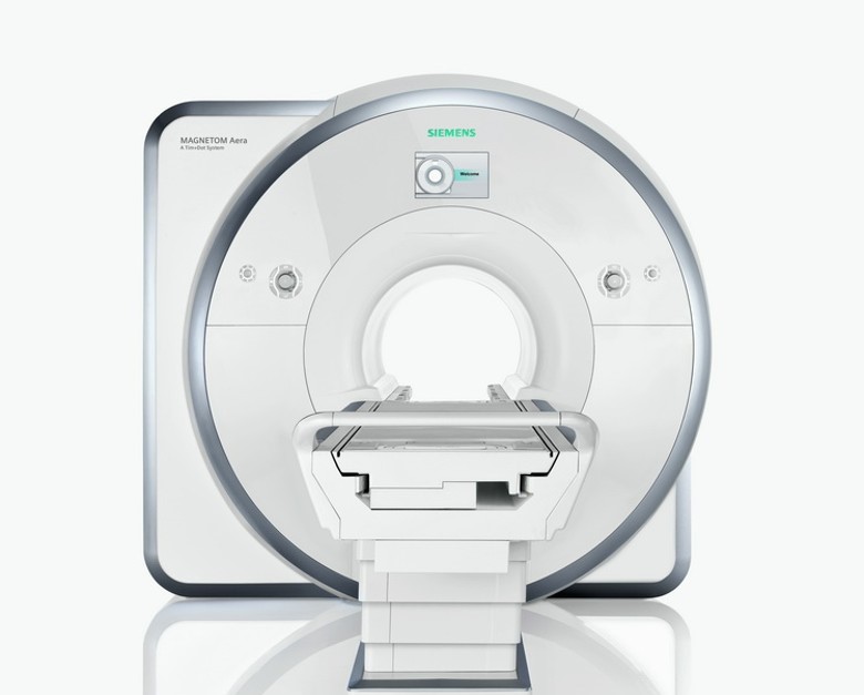
Magnetic Resonance Imaging (MRI)
Magnetic resonance scans are non-invasive tests that use high magnetic fields and radio waves to display detailed images of the inside of the human body for tumor assessment and detection of cancer causes. Magnetic resonance scanning is often used to evaluate the whole body, heart, brain and other parts. Because there is no radiation, it is suitable for physical examination, evaluation and follow-up in most parts of the human body (including the musculoskeletal system and nervous system)
MRI works by using a powerful magnetic field and radio waves to stimulate hydrogen nuclei (protons) in the body, which emit signals that are processed by a computer to create an image. As water molecules contain protons and the human body consists mostly of water, MRI is a useful diagnostic tool for examining the body.

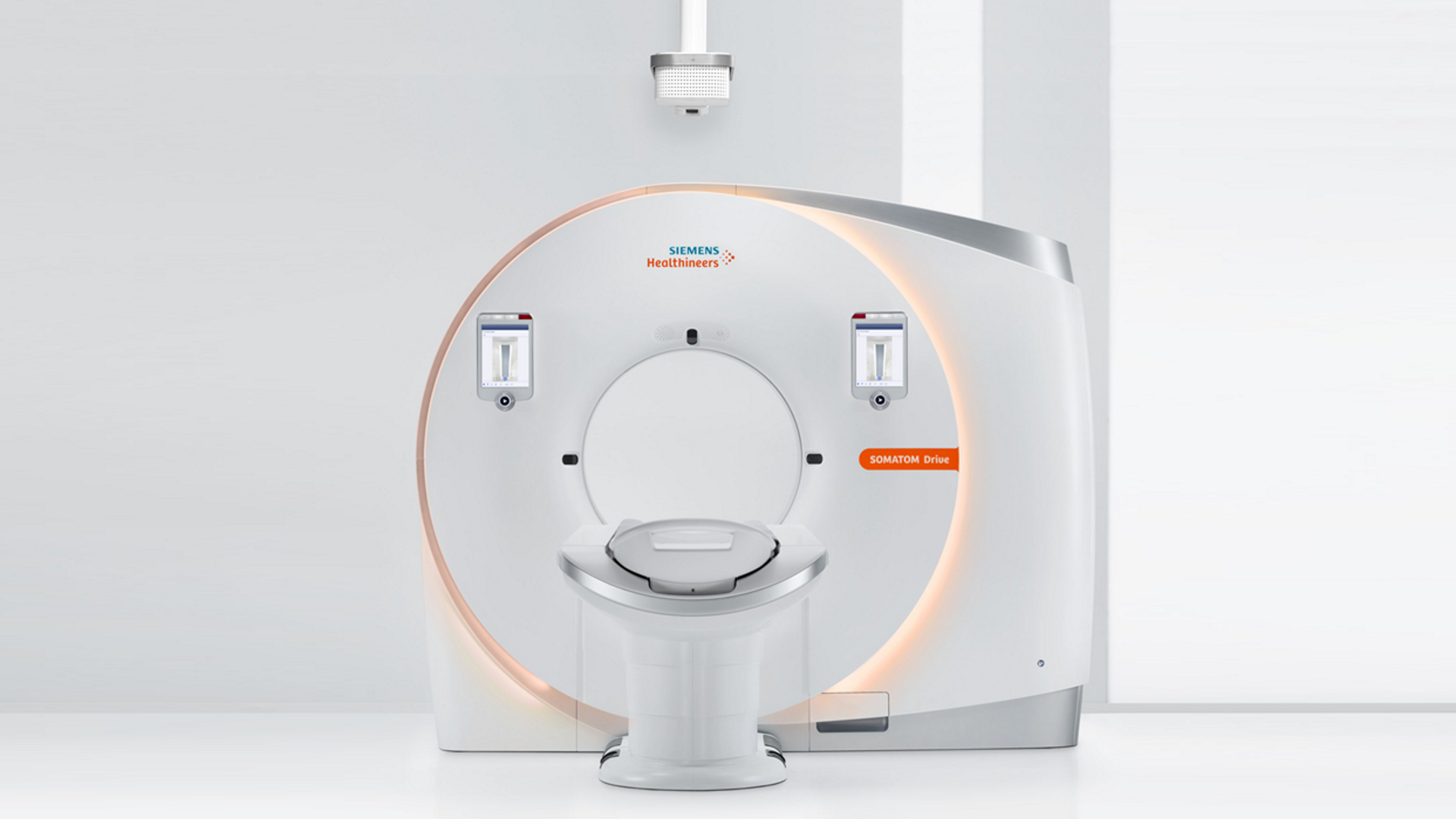
Computed Tomography (CT) Scan
Computer scanning uses X-rays to project around the object from multiple angles. The computer analyzes the image data to produce fine cross-sectional images to observe the internal structure of the object. It is widely applicable to different body parts. This is one of the most commonly used medical imaging technologies. Computer scans are commonly used to evaluate infectious diseases, cardiovascular diseases, musculoskeletal diseases, tumors and cancers.

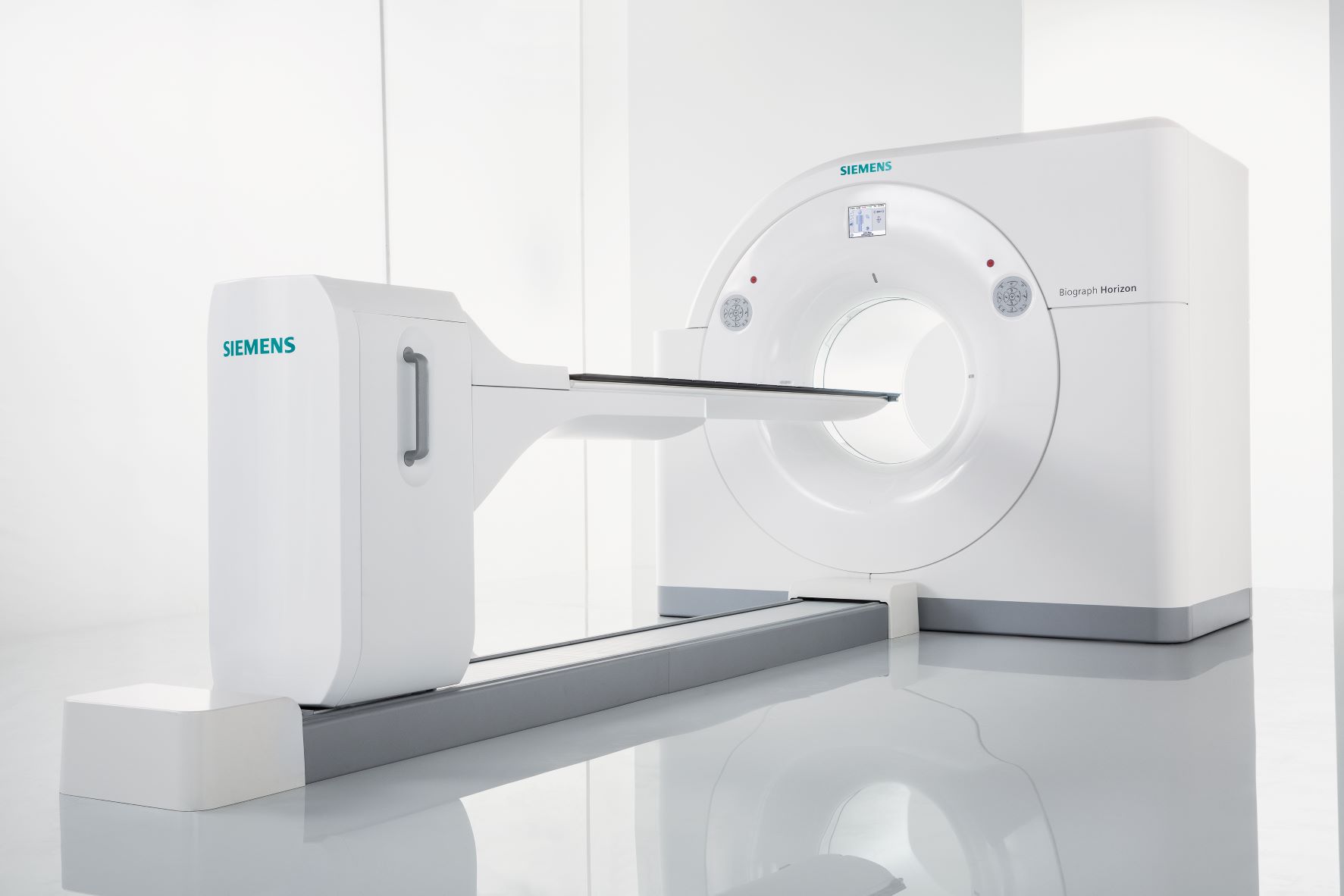
Positron Emission Tomography (PET) Scan
Positron emission scans are often used to evaluate and evaluate cancer, malignant tumor location, brain nervous system disorders, and cardiovascular diseases. It is a functional medical imaging examination that can provide detailed information about the function of a certain body organ or system. Before the scan, the radiologist will inject radioactive isotope medicine into the patient's body. During the scanning process, a special camera will detect the amount of radiation released by the drug, and then a computer will construct a multi-dimensional image of the inspected area. Radioisotope drugs often accumulate in diseased tissue rather than healthy tissue.
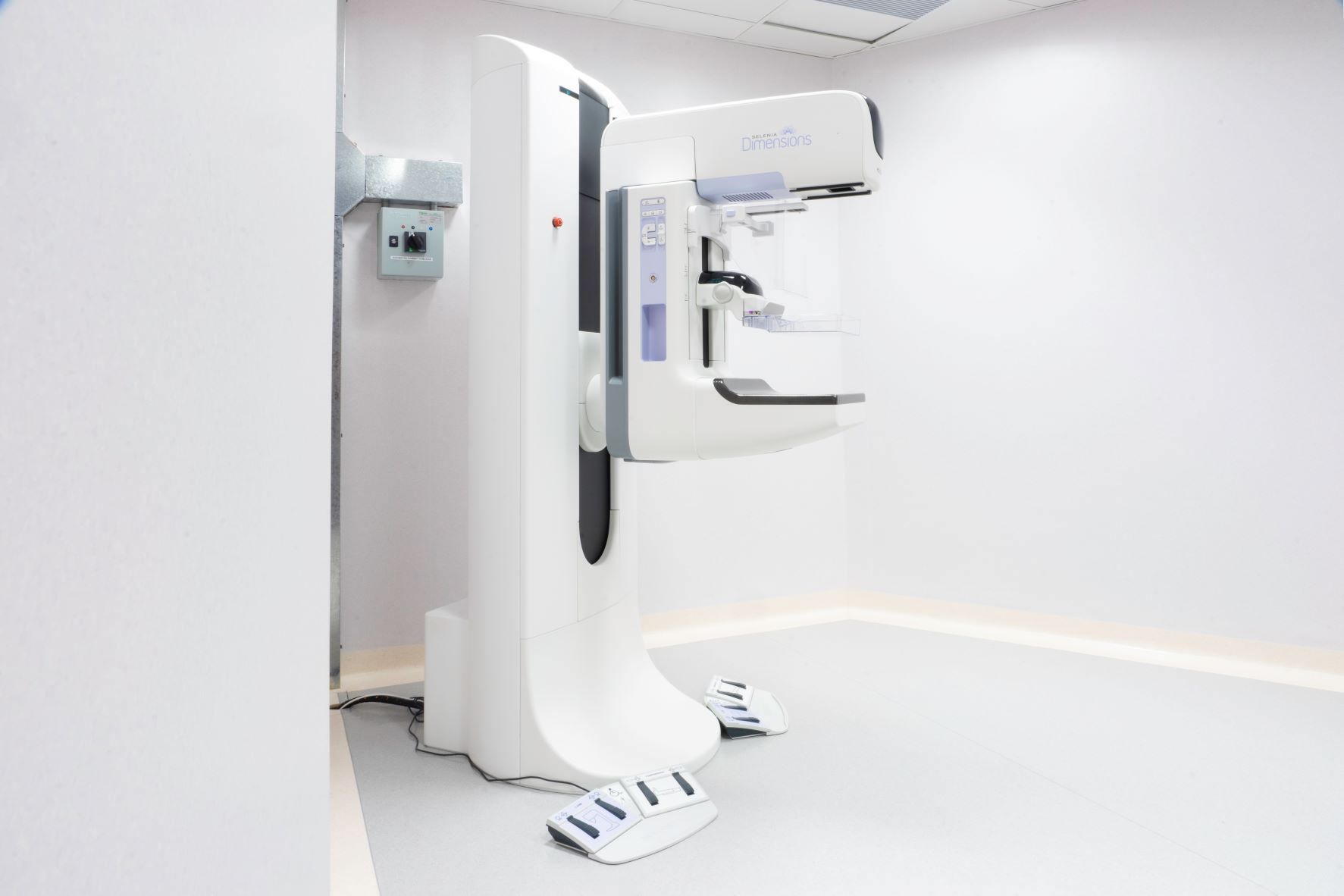
3D Mammography
3D mammography was approved by the U.S. Food and Drug Administration in 2011 and is a brand-new breast imaging technology. 3D mammography produces multiple low-dose X-rays of the breast from different angles to create a set of 3D images. Traditional mammography only obtains 2D images of the breast from two different angles. 3D mammography is used to evaluate early pathological changes in the breast, such as cysts or tumors - earlier than the patient notices. By examining the 3D reconstructed breast images layer by layer, radiologists can make assessments more easily.

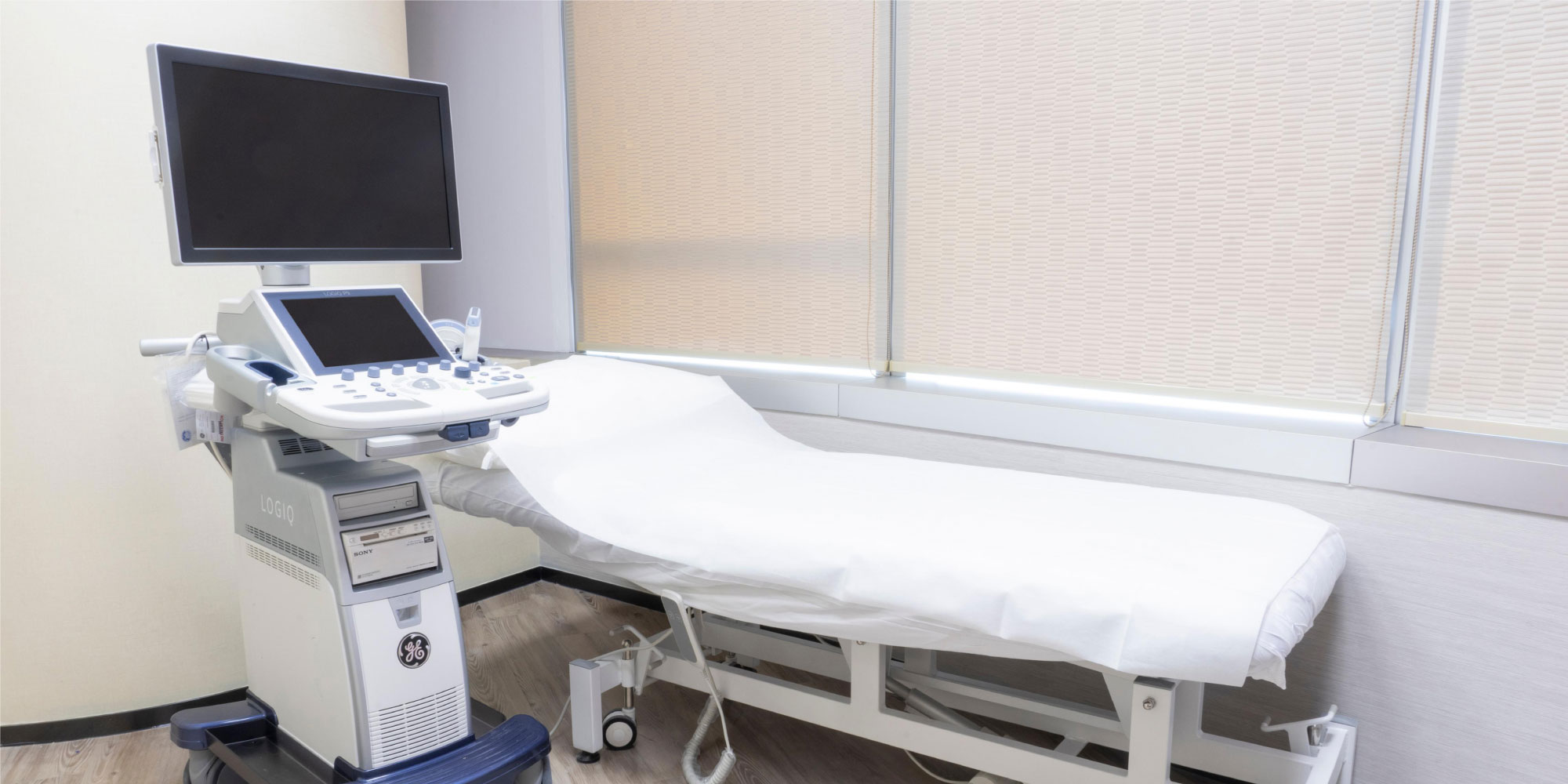
Ultrasound Examination
Ultrasound imaging uses high-frequency sound waves to produce real-time images of the inside of the body. It involves using a small transducer (probe) and ultrasound gel placed directly on the skin. High-frequency sound waves travel from the probe through the gel into the body. A transducer collects the bounced sound, which is then run through a computer to create an image. Ultrasound examination is often used to examine body organs such as the uterus, pelvis, liver, kidneys, etc. It can help evaluate diseases of different organs, examine the baby of a pregnant woman, and examine the baby's brain and buttocks. It is also used to help guide biopsies and evaluate heart disease. Ultrasound is safe, painless, non-invasive, and unlike X-rays and CT scans, does not use ionizing radiation.
Ultrasound imaging displays real-time images of internal organs, blood flow through vessels, and fetal activity in the uterus.
Ultrasound technology has advanced to produce three-dimensional (3D) images from two-dimensional (2D) images, while Doppler ultrasound allows radiologists to visualize and assess blood flow in arteries and veins throughout the body, including various organs and regions such as the abdomen, arms, legs, neck, and brain (in infants and children).
Related Information
To provide advanced, accurate, and efficient MRI scans, we use the Siemens MAGNETOM Aera 1.5T MRI scanner, a high-tech device that utilizes Tim 4G technology to improve image quality and speed, and the Dot engine to enhance the accuracy, repeatability, and efficiency of the examination process. In addition, the Aera features a 70 cm-wide open bore design and a total length of only 145 cm, which increases patient comfort and reduces the likelihood of claustrophobia during certain examinations. The new technology also reduces noise levels by up to 70%, further optimizing the examination environment.
The Siemens SOMATOM Drive is one of the most advanced dual-source computer scanners available, and there are currently only three of them in Hong Kong.
Dual-source computer scanners are equipped with two sets of X-ray tubes and detectors, which move around the patient to capture images. This system enables the scanners to capture images at twice the speed of traditional single-source computer scanners.
The SOMATOM Drive scanner incorporates advanced technology that enhances precision, efficiency, and patient safety while maintaining high-quality images. Notably, it reduces motion artifacts and respiratory motion through ultra-fast scan times, low radiation dose, and minimal contrast agent usage. These advancements not only improve the patient experience but also have many applications, including cardiovascular imaging, functional imaging, pediatric imaging, and geriatric imaging.
Positron emission tomography (PET) itself cannot provide accurate location information, meaning it cannot correlate the radioactive isotope signals received from the patient's body to the precise location of the organs or tissues emitting the isotopes. However, by combining this with computed tomography (CT) technology, which integrates the patient's anatomical structure information with the physiological function data provided by the PET scan, it is possible to accurately clarify the issues at hand. This allows doctors to accurately understand the condition and design subsequent treatment plans.
Better Detection. Faster. Less Radiation.
The 3D Hologic Selenia Dimensions technology provides exceptionally clear breast images. This instrument detects an average of 41% more invasive breast cancers than traditional 2D mammography, reducing false positive cancer results by up to 40%. The newly improved technology even reduces breast compression time to 3.7 seconds, and the machine's ergonomic design significantly improves patient comfort. This revolutionary system is the world's first and only mammography instrument capable of guiding a biopsy, and it is approved by the U.S. Food and Drug Administration for image-guided vacuum-assisted biopsy. The radiation dose of this 3D mammography is similar to that of traditional 2D mammography; the C-view software synthesizes 2D images from tomographic data, further reducing the patient's radiation dose.
The Hong Kong Breast Cancer Foundation recommends routine breast imaging screenings every 2 years for women aged 40 and above. If you have breast cancer symptoms or a strong family history ofMammary Cancer, seek medical assistance and schedule a breast imaging screening. Early detection can improve the chances of successful treatment.
Hotline
-
Telephone Inquiry
New customer inquiry: 3001 5810
Existing customers: 2156 5838 -
WhatsApp inquiry
3001 5810 -
E-mail
enquiry@reviveonco.com.hk -
Opening Hours
Mon-Fri: 9:30am - 6:30pm
Sat: 9:30am - 1:30pm
Closed on Sundays and Hong Kong public holidays -
Mong Kok Clinic
12/F, Office Tower, Langham Place, 8 Argyle Street, Mong Kok, Kowloon
Fax: 3579 0153
EC Specialists Premium (PHF No.: DP000104)